
3.2 Leaves
We see a massive amount of variation in the sizes and shapes of leaves. Similarly, the anatomical structure of leaves can vary considerably. Plant leaves may be specialised to maximise light utilisation, to minimise water loss, to facilitate C4 photosynthesis or CAM photosynthesis, to resist damage due to water stress, or to float on water.
Media Attributions
- 3.8. Cuticle upper epidermis © Berkshire Community College Bioscience Image Library is licensed under a Public Domain license
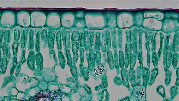
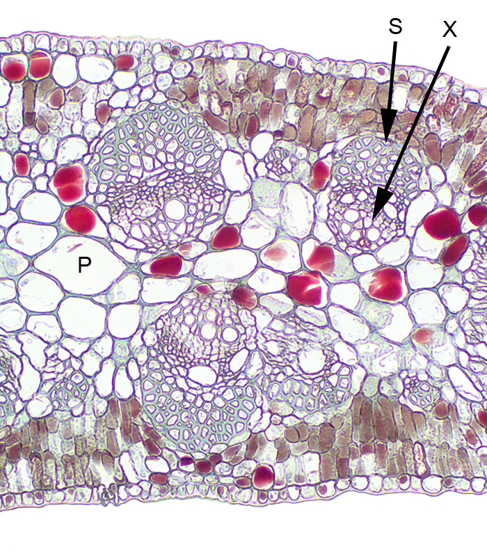
- Figure 3.9. Mesophyte Syringa leaf © Berkshire Community College Bioscience Image Library is licensed under a Public Domain license
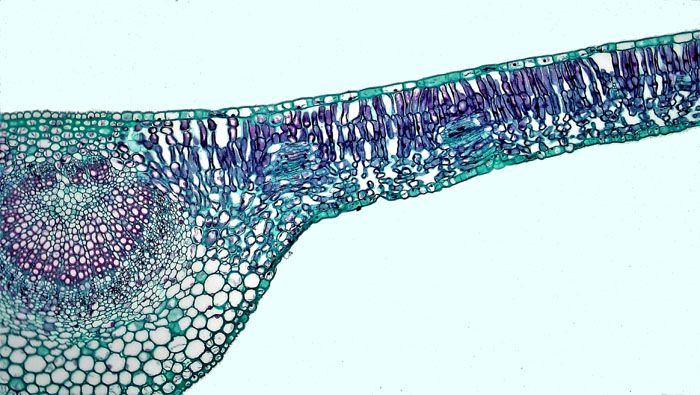
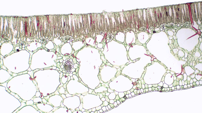
- 3.10. Xerophyte rhizopora © Sean Bellairs is licensed under a CC BY-SA (Attribution ShareAlike) license
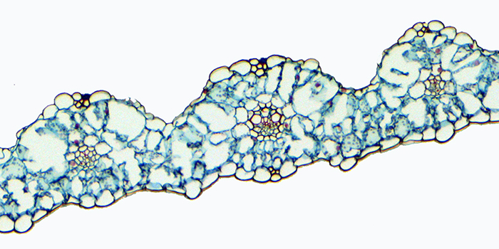
- 3.11. Callistemon leaf © Sean Bellairs is licensed under a CC BY-SA (Attribution ShareAlike) license
- 3.12. Acacia 3 © Sean Bellairs is licensed under a CC BY-SA (Attribution ShareAlike) license
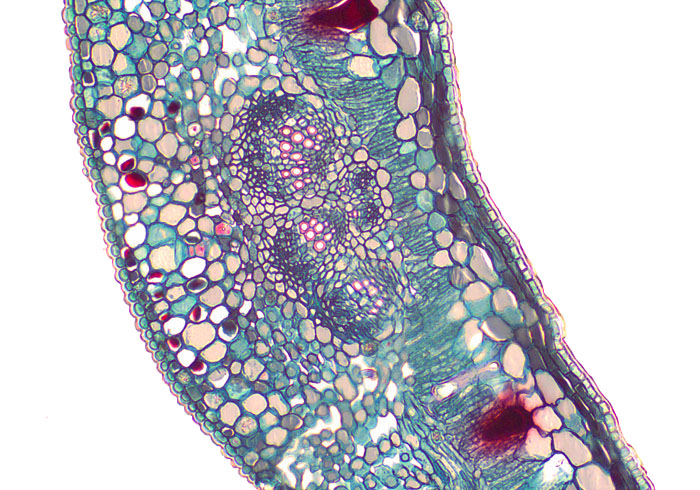
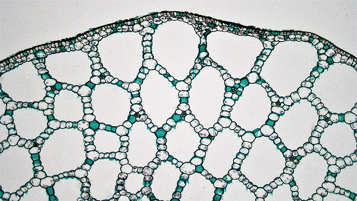
- 3.13. Hydrophyte nymphaea © Sean Bellairs is licensed under a CC BY-SA (Attribution ShareAlike) license
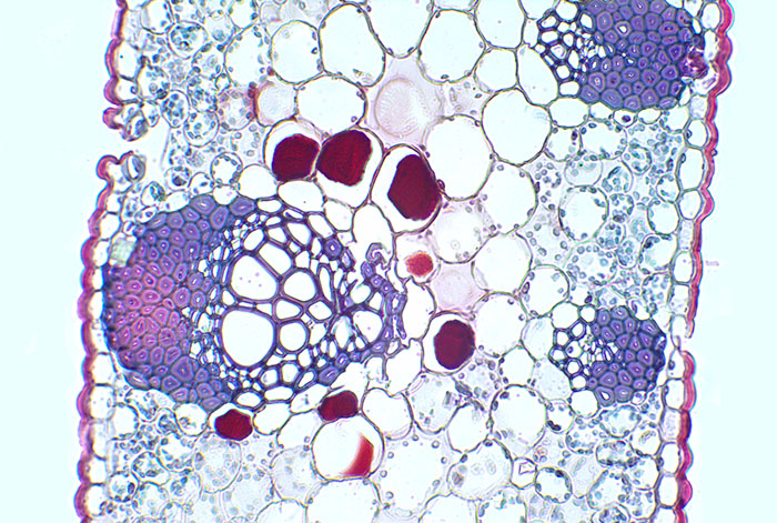
- 3.14 Aerenchyma in Potamogeton © Berkshire Community College Bioscience Image Library is licensed under a Public Domain license
- 3.15.a. Wheat leaf © BlueRidgeKitties is licensed under a CC BY-NC-SA (Attribution NonCommercial ShareAlike) license
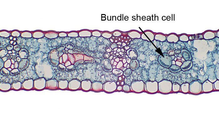
- 3.15.b. Zea mays bundle sheath cells © Sean Bellairs is licensed under a CC BY-SA (Attribution ShareAlike) license
