
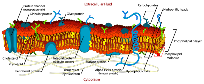
1.3 Cell membrane
Immediately inside the cell wall there is a cell membrane surrounding the contents of the cell. It is made up of a phospholipid bilayer with proteins. The outer surface of the phospholipid layer is attracted by water (hydrophilic) whereas the tail of the phospholipid molecule is repelled by water (hydrophobic). The proteins may be embedded in the membrane or just attached to the surface. Some of the embedded protein molecules pass right through the membrane and are important for transport of substances through the membrane.
1.3.1 Cell Membrane functioning
The cell membrane is important for compartmentalising different parts of the cell to allow metabolic functioning to occur and to control substances in the cell. It allows control of substances entering the cell and it allows the cell to compartmentalise waste into the vacuole. Oil droplets, vesicles from the golgi apparatus and vacuoles just have a single bilipid membrane around them. The nucleus, chloroplasts and mitochondria have a double bilipid membrane around them.
1.3.2 Diffusion
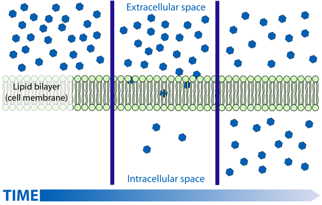
Diffusion is the process of random movement of molecules towards a state of equilibrium; the net movement is always from the direction of greater concentration to lesser concentration and in complex solutions, each substance moves independently of the other. Substances tend to diffuse until they are evenly distributed.
Small, non-polar molecules can pass through the lipid bilayer of a membrane by diffusing through it.
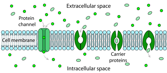
1.3.3 Facilitated diffusion
Facilitated diffusion also involves molecule movement down a concentration gradient until the concentration of molecules are equal on both sides of the membrane. However in this case the solute molecules do not move through the membrane on their own. They combine with a carrier molecule in the membrane which allows the solute molecule to pass through the membrane. No energy is used and they pass through the carrier protein until the concentration is the same on both sides of the membrane.
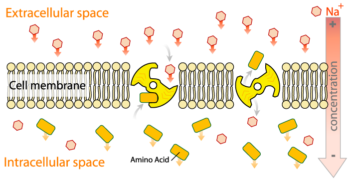
1.3.4 Active transport
Active transport is different from the other transport processes above in that it involves transport of a solute against a concentration gradient (i.e. from an area of low concentration to one of higher concentration). This process relies on carrier molecules but also requires energy as it forces molecules to move against the concentration gradient. Energy comes from the energy storage molecule ATP and is generated through cellular respiration.
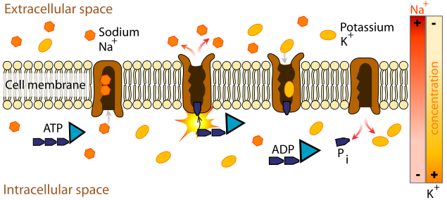
Active transport may also be indirect. In the figure below the sodium ions have been concentrated above the membrane. The sodium ions seek to equilibrate the concentration each side of the membrane but for the sodium ions to travel though the membrane, the protein carrier requires an amino acid be attached and pumped in the opposite direction. Thus actively concentrating the sodium ions then results in transport of the amino acids indirectly as the sodium ions move down the concentration gradient.
Media Attributions
- Figure 1.6. The cell membrane of a plant cell © Mariana Ruiz adapted by Alokprasad84 is licensed under a CC BY (Attribution) license
- Figure 1.7. A diagram showing diffusion of a small non-polar molecule through a membrane © Mariana Ruiz Villarreal is licensed under a Public Domain license
- Figure 1.8. A diagram showing a molecule passing through a membrane passively using protein channels or carrier proteins © Mariana Ruiz Villarreal is licensed under a Public Domain license
- Figure 1.9. Sodium ions passing through a membrane © Mariana Ruiz Villarreal is licensed under a Public Domain license
- Figure 1.10. Indirect active transport due to a active transport achieving a high sodium concentration © Mariana Ruiz Villarreal is licensed under a Public Domain license
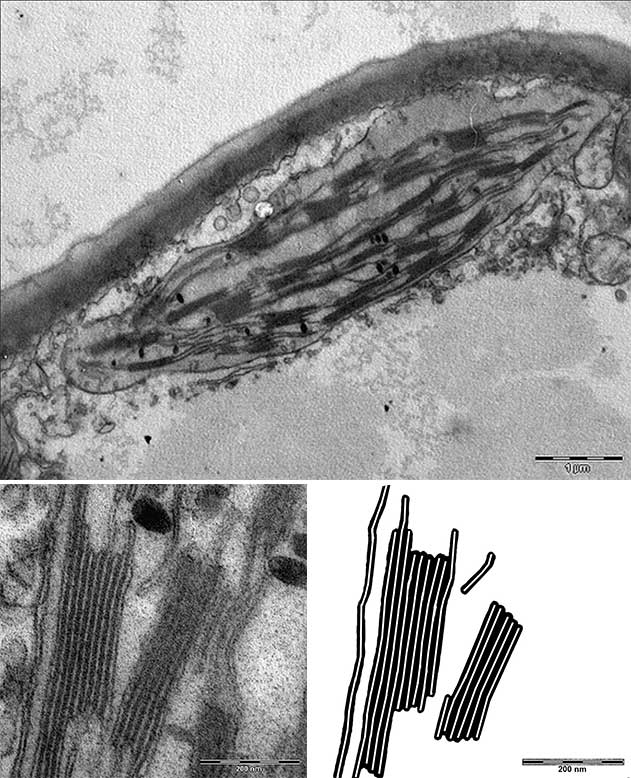
- Figure 1.11. Membrane structures in plants © Bela Hausmann adapted by Sean Bellairs is licensed under a CC BY-SA (Attribution ShareAlike) license
