
3.1 Stems
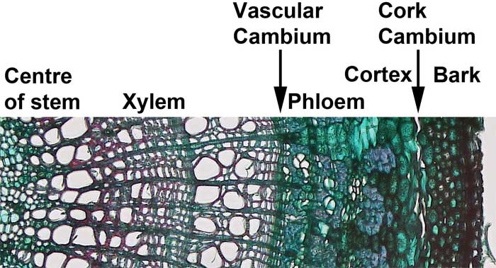
Stems are produced by the primary apical meristem in but may be increased in girth in woody plants due to secondary growth. Secondary growth is produced by lateral meristems in the woody stems and roots of woody plants.
Secondary xylem and secondary phloem are produced from a cylinder of meristematic tissue within the woody stems and roots. This cylinder of meristematic tissue is the vascular cambium. The secondary xylem provides additional structural support and additional water conduction tissue in shrubs and trees. The secondary phloem replaces the primary phloem.
Similarly, as the trunk of a woody plant gets larger, the dermal tissue need to be expanded and replaced. New dermal tissue is produced by the cork cambium, which lies beneath the bark.
Media Attributions
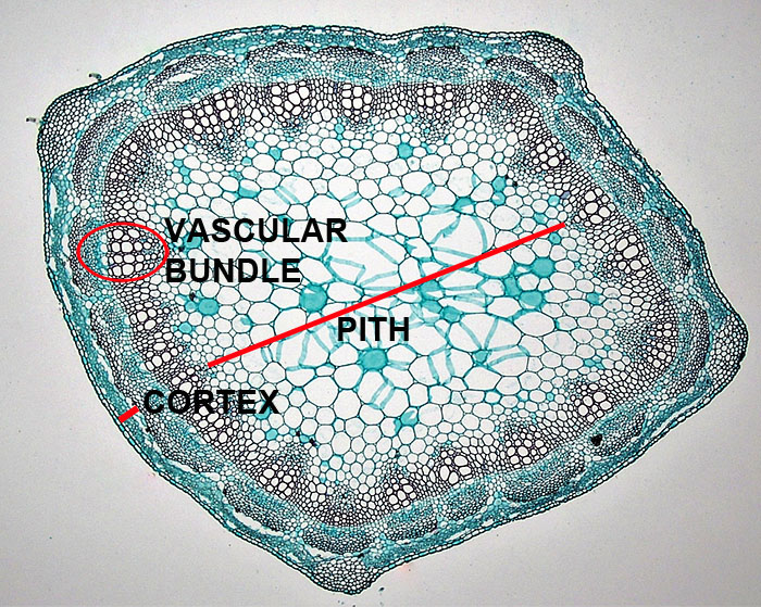
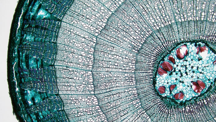
- Figure 3.1 Trifolium primary stem © Berkshire Community College Bioscience Image Library adapted by Sean Bellairs is licensed under a CC BY-SA (Attribution ShareAlike) license
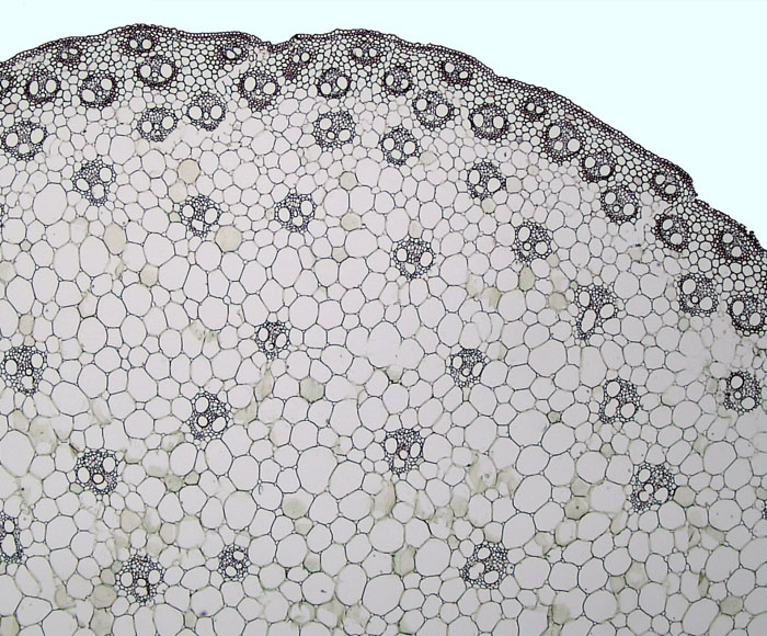
- Figure 3.2. Zea mays stem © Berkshire Community College Bioscience Image Library adapted by Sean Bellairs is licensed under a CC BY-SA (Attribution ShareAlike) license
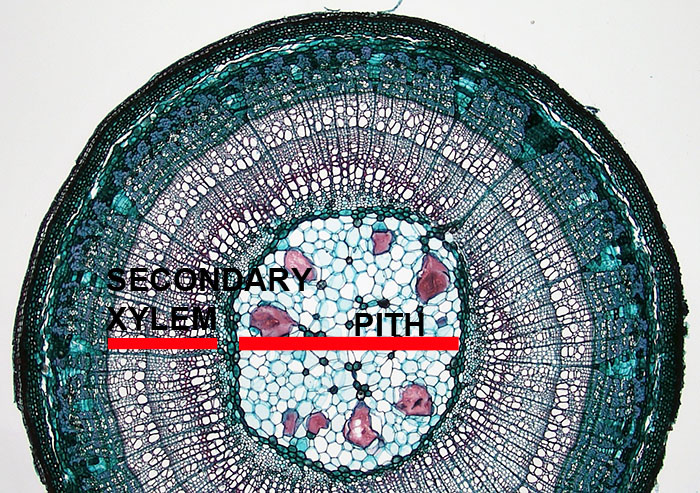
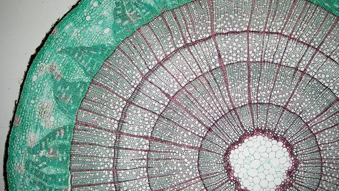
- Figure 3.3. Two year Tilia © Berkshire Community College Bioscience Image Library adapted by Sean Bellairs is licensed under a CC BY-SA (Attribution ShareAlike) license
- Figure 3.4. Vascular cambium © Berkshire Community College Bioscience Image Library adapted by Sean Bellairs is licensed under a CC BY-SA (Attribution ShareAlike) license
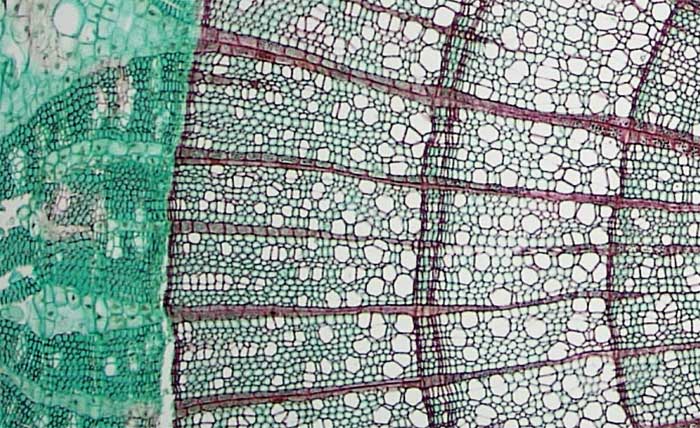
- Figure 3.5. Tilia 3 growth rings © Berkshire Community College Bioscience Image Library adapted by Sean Bellairs is licensed under a CC BY-SA (Attribution ShareAlike) license
- Figure 3.6. Liriodendron stem © Berkshire Community College Bioscience Image Library is licensed under a Public Domain license
- Figure 3.7. Liriodendron stem © Berkshire Community College Bioscience Image Library adapted by Sean Bellairs is licensed under a CC BY-SA (Attribution ShareAlike) license
