
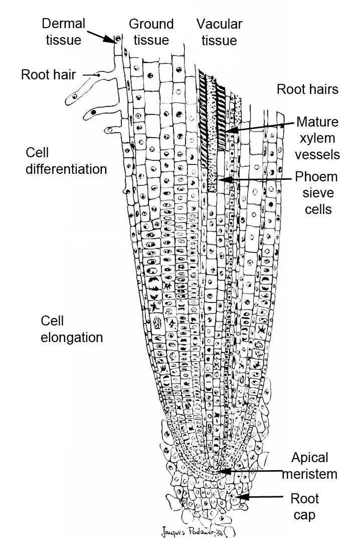
3.3 Roots
Though unseen, the roots of a plant also have specialist anatomical features that enable plants to efficiently obtain nutrients and control the substances entering a plant.
Media Attributions
- Figure 3.16 Root structure © Douglas Marsland adapted by Sean Bellairs is licensed under a CC BY-SA (Attribution ShareAlike) license
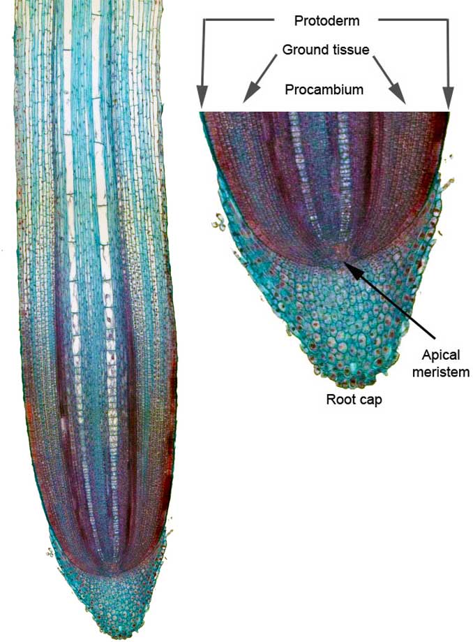
- Figure 3.17. Zea mays root tip © Jen Dixon adapted by Sean Bellairs is licensed under a CC BY-SA (Attribution ShareAlike) license
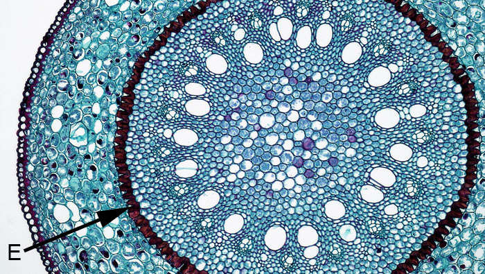
- 3.18. Smilax root with endodermis © Berkshire Community College Bioscience Image Library adapted by Sean Bellairs is licensed under a CC BY-SA (Attribution ShareAlike) license
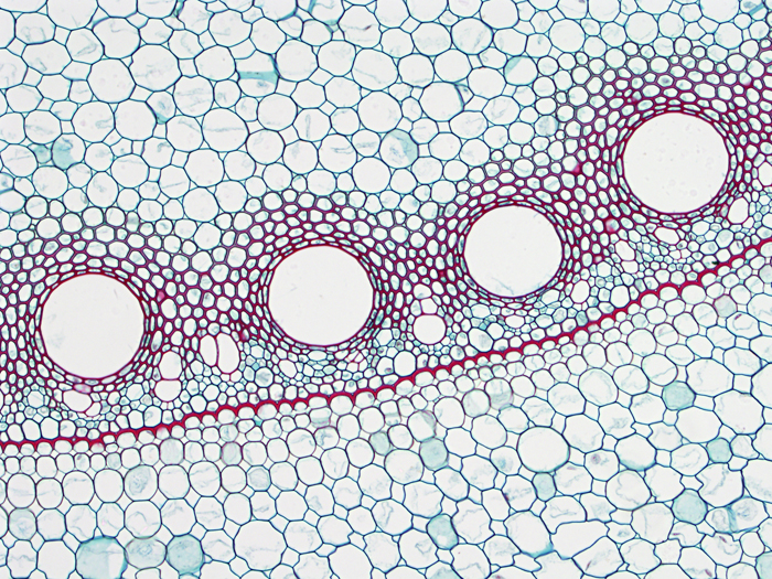
- 3.20. Zea mays endodermis © BlueRidgeKitties is licensed under a CC BY-NC-SA (Attribution NonCommercial ShareAlike) license
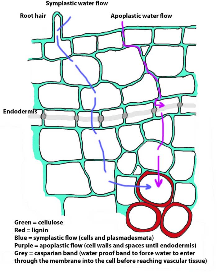
- 3.20. Water movement through root © Sean Bellairs is licensed under a CC BY-SA (Attribution ShareAlike) license
